How useful is MRI to diagnose spondyloarthritis?

Just a quick thought regarding the use of MRI for inflammatory spinal pain.
MRIs are often thought of as the "gold standard" to aid diagnosis in spinal pain.
MRIs of the spine are being used to exclude the presence of serious pathology such as malignancies. They are used to look for spinal cord or nerve root compression.
I order a lot of MRIs of the spine, probably due to the easy access where I work. I find them useful for some mechanical spinal disease and particularly useful in the assessment of chronic spinal pain which has features suspicious for inflammatory spinal disease (read Take this test: Do you have Ankylosing Spondylitis?).
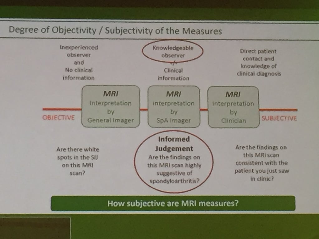
I share this slide to highlight that MRI interpretation is not black and white. There is a subjective component to it as it's not a perfect test.  "Slide by Robert Lambert, Radiologist, presenting at the 10th International Congress on Spondyloarthritides"
What this means is that a "negative" MRI with the correct settings for inflammatory disease may not necessarily rule out inflammatory spinal pain.
In addition, a "positive" MRI depending on the degree of certainty or the precision of the reporting may not actually be definitive for the diagnosis.
Factors which may influence what is reported on the MRI include the actual stage of the disease (i.e. very early vs later disease) and the experience of the reporting radiologist in looking at these sort of MRIs in these patient groups.
Your rheumatologist is a detective and an expert diagnostician. This involves collecting the clues from a good history, followed by an examination, and then reconciling this with the "objective" findings on investigations such as the MRI.
We rheumatologists do not practice an exact science all the time, and with spondyloarthritis, experience and intuition, with educated guesswork plays a part.


