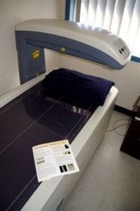
Not all DXA scans are created equal

 By Suzy Oglesby, Exercise Physiologist & DXA technician
Seeking the “best” is something that we as humans value. It holds true for results, new purchases and of course it also applies to the health care industry. While many patients will brag about seeing the “best” highly regarded specialists, little attention is paid to the experience and reputation of those technicians who perform imaging services.
It is surprising really as we often rely heavily on such modalities to confirm or shed light on a diagnosis. Many studies often acknowledge the specific technology-related error of such imaging techniques, however inter and intra-tester reliability is often overlooked.
As a DXA (dual-energy X-ray absorptiometry) technician, I don’t take my job lightly.
DXA is used to diagnose Osteoporosis.
I know that a poorly taken scan can potentially change the clinical diagnosis that is based on these results. Luckily there are steps that can be taken as I strive to be the best and more importantly to ensure that the results from my scans are accurate and reliable.
The first step involves a daily quality assurance check of the DXA machine via a series of tests and also using a “phantom” spine which is simply a mock spine used as a control. A measurement is recorded for the phantom and then compared to the database of previous results to ensure that it sits within the small acceptable range. Once this is done, I know that the accuracy of the day’s scans rest firmly upon my shoulders and the steps that I take to minimize error.
So what constitutes a poorly taken scan?
A DXA scan can be significantly affected by the positioning of the patient, any movement during the scan, and by artifact such as coins, metal zippers and buttons. Ensuring that the appropriate body part thickness is selected is also required. These are factors that the technician must aim to control for to optimize results.
Once the scan is obtained, a trained eye is necessary to ensure that analysis itself is accurate. While the computer will automatically identify the region for analysis, most people have at some stage experienced the frustration of technology and can vouch for the fact that these amazing machines don’t always get it right!
In fact, there is rarely a scan that is done that I don’t have to alter in some way. Bone density and body composition measurements rely on the appropriate allocation of tissue type for accurate analysis and hence these small alterations can have a large bearing on the result.
In addition to the daily routine, DXA technicians also perform tests of precision error whereby numerous repeat scans are taken and assessed for variation. Acceptable values are between 1-2% for the spine and 2-3% for the hip.
As is the case for most professions, greater experience leads to better results and this is certainly the case for DXA. Experience allows the technician to develop and refine their skills to detect any abnormalities, further reducing the potential for error.
The more “normal” scans that you see, the more obvious the “abnormal” scans are.
Having an imaging technician who is conscious of minimizing error means that as a patient you can be confident in any decisions that are made based on the results of the technique.
